Home

غلاديس فهرنهايت قابس كهرباء mitochondria light microscope صريح اختياري اعتزم

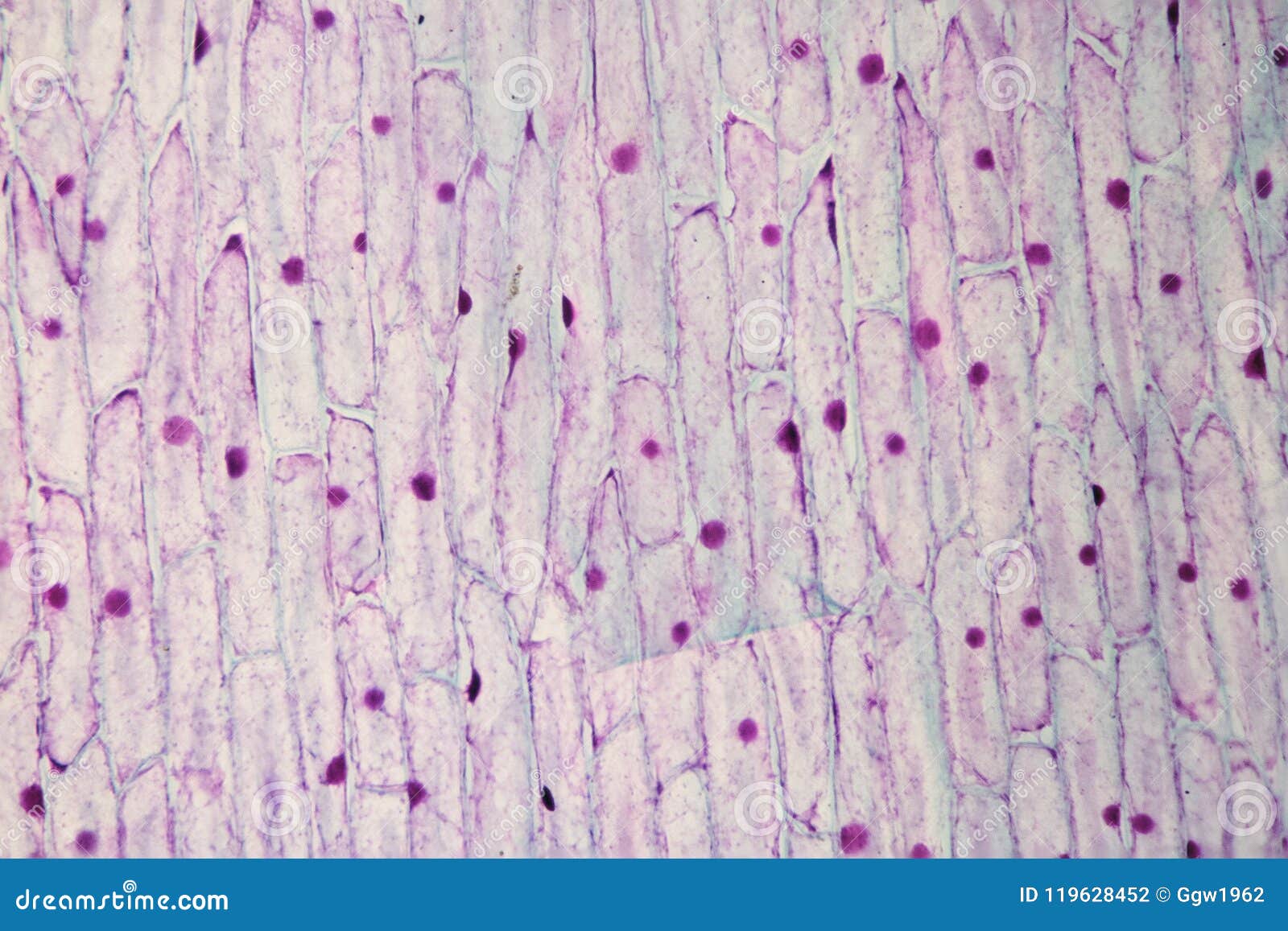
Plant mitochondria stock photo. Image of energy, research - 119628452

A method for freeze-fracture and scanning electron microscopy of isolated  mitochondria - ScienceDirect
A method for freeze-fracture and scanning electron microscopy of isolated mitochondria - ScienceDirect

51 Make: mitochondria ideas | mitochondria, mitochondrial, mitochondrial  disease
51 Make: mitochondria ideas | mitochondria, mitochondrial, mitochondrial disease

Light microscopic methods to visualize mitochondria on tissue sections -  ScienceDirect
Light microscopic methods to visualize mitochondria on tissue sections - ScienceDirect

Plant Mitochondria Viewed Under The Microscope. Stock Photo, Picture And  Royalty Free Image. Image 118494415.
Plant Mitochondria Viewed Under The Microscope. Stock Photo, Picture And Royalty Free Image. Image 118494415.

Cell Image Library
Cell Image Library

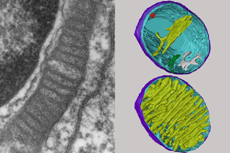
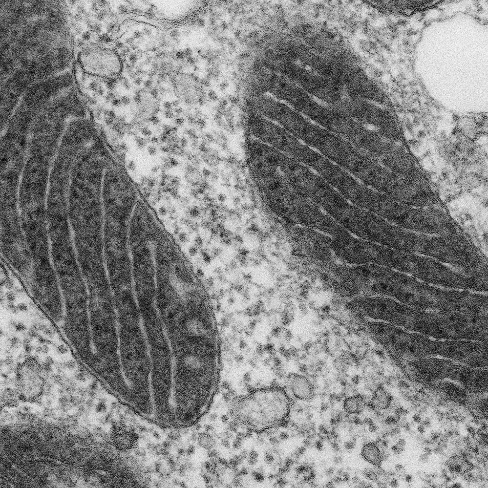
Insight into mitochondrial structure and function from electron tomography  - ScienceDirect
Insight into mitochondrial structure and function from electron tomography - ScienceDirect

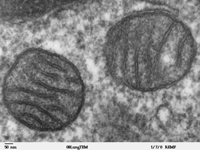
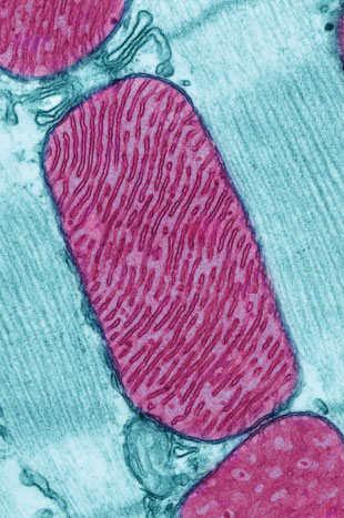
Mitochondrion - Wikipedia

Mitochondria under the microscope — Science Learning Hub

Pin on Stories
Pin on Stories

Wellcome Trust Centre for Mitochondrial Research Newcastle UK

Microscopes
Microscopes

Can Red Light Therapy Aid Mitochondria for ATP Production in Cells?

Are mitochondria the natural cellular light sources?

When we see a cheek cell through a microscope, can we see the mitochondria?  - Quora
When we see a cheek cell through a microscope, can we see the mitochondria? - Quora

Mitochondria - The School of Biomedical Sciences Wiki

Live mitochondria seen in unprecedented detail: photobleaching in STED  microscopy overcome
Live mitochondria seen in unprecedented detail: photobleaching in STED microscopy overcome

7 Things To Know About Mitochondria: 2016 edition

Light and electron microscopy showing ultrastructural changes in the... |  Download Scientific Diagram
Light and electron microscopy showing ultrastructural changes in the... | Download Scientific Diagram
Natural History Magazine

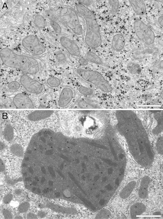
Three-dimensional ultrastructure of giant mitochondria in human  non-alcoholic fatty liver disease | Scientific Reports
Three-dimensional ultrastructure of giant mitochondria in human non-alcoholic fatty liver disease | Scientific Reports

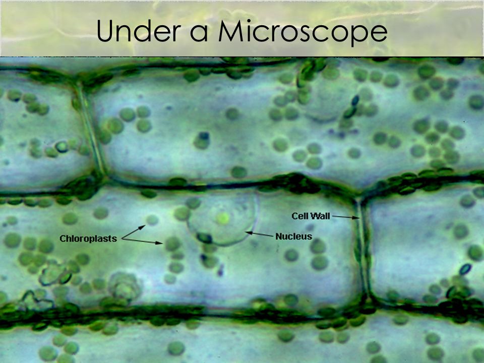
Can you expect to see mitochondria while using a light compound microscope?  | Socratic
Can you expect to see mitochondria while using a light compound microscope? | Socratic

Light microscopy Images of clusters of isolated mitochondria on mica at...  | Download Scientific Diagram
Light microscopy Images of clusters of isolated mitochondria on mica at... | Download Scientific Diagram

A new glow for electron microscopy | MIT News | Massachusetts Institute of  Technology
A new glow for electron microscopy | MIT News | Massachusetts Institute of Technology

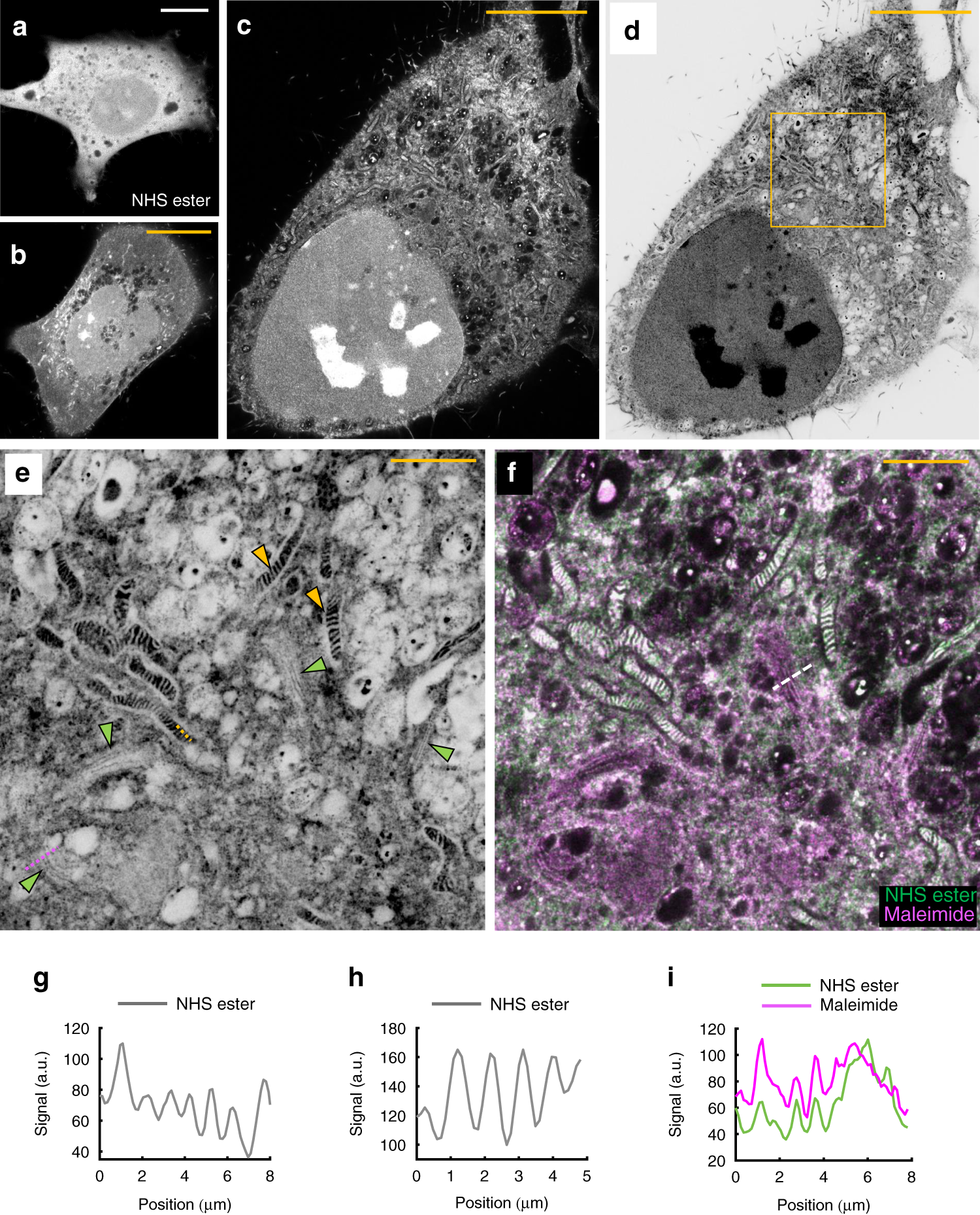
Light microscopy of proteins in their ultrastructural context | Nature  Communications
Light microscopy of proteins in their ultrastructural context | Nature Communications

Light and electron microscopy of mitochondria in the oocytes of Argulus...  | Download Scientific Diagram
Light and electron microscopy of mitochondria in the oocytes of Argulus... | Download Scientific Diagram

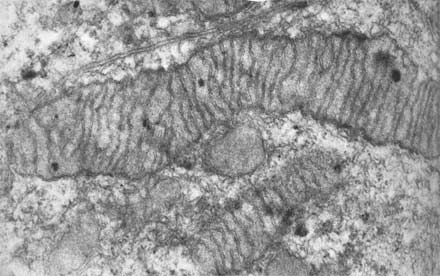
Cytology. Cytology. rods, or threads which vary in size, number, and  distribution from cell to cell (Figure 3-2 (a) and (b) ). They are most  readily identified in the living cell
Cytology. Cytology. rods, or threads which vary in size, number, and distribution from cell to cell (Figure 3-2 (a) and (b) ). They are most readily identified in the living cell

The Shrinking Mitochondrion | The Scientist Magazine®

Cell Lab
Cell Lab